THE HIGHEST QUALITY DIGITAL X-RAY, DR FLAT PANEL, OR X-RAY EQUIPMENT AT THE LOWEST PRICE GUARANTEED.
XRAY Solutions Digital X-ray Machines and Equipment
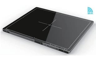
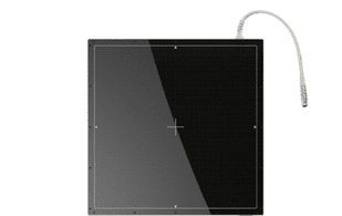
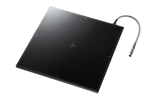
Welcome to XRAY Solutions, your one-stop shop for high-quality, affordable digital X-ray DR Flat Panel Detectors and X-ray equipment! We’re dedicated to providing healthcare professionals with the cutting-edge tools they need to deliver accurate diagnoses and exceptional patient care.
Why Choose X-RAY Solutions?
Unmatched Quality: We partner with industry-leading manufacturers to bring you the most reliable and advanced digital X-ray systems available.
Affordability: We understand the financial pressures faced by healthcare practices. That’s why we offer a wide range of X-ray machines and equipment in competitive prices and flexible financing options to fit your budget.
Specialization: We have a deep understanding of the unique needs of chiropractic, veterinary, podiatry, urgent care, and medical practices. Our team of experts will work closely with you to find the perfect digital X-ray systems for your specific requirements.
Exceptional Service: We’re committed to providing you with the highest level of customer service. Our experienced team is available 24/7 to answer your questions and support you every step of the way.
Our Specialities
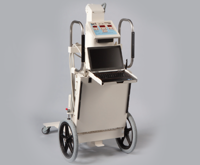
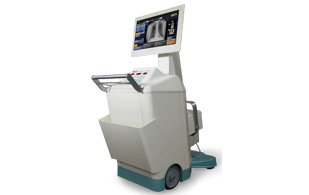
Mobile and Portable X-ray: Respond quickly and effectively with our fast-deployment mobile portable X-ray units. Capture immediate images for on-the-spot diagnoses and streamlined patient care in your urgent care setting.
Podiatry: Get detailed views of feet and ankles with our DR panels featuring exceptional soft tissue clarity. Our systems support weight-bearing imaging, crucial for accurate podiatric assessments.
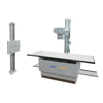
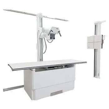
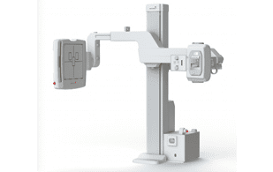
Medical Practices: Elevate your diagnostic capabilities with our comprehensive range of DR flat panels, generators, and C-arms for X-ray room systems. We offer scalable solutions, from compact systems for individual offices to robust configurations for larger medical facilities.
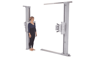
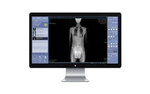
Chiropractic: Enhance posture analysis and spinal alignment assessments with high-resolution DR flat panels designed for precise skeletal visualization. Our compact and flexible chiropractic X-ray systems fit seamlessly into your space.
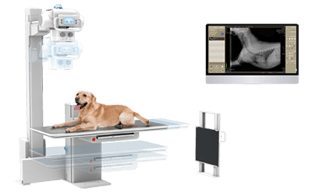
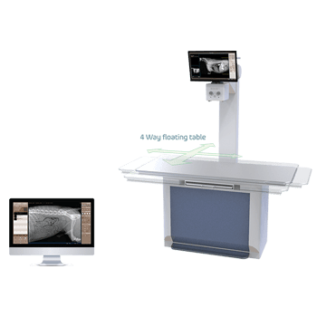
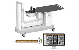
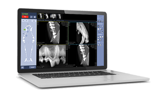
Veterinary: Capture clear images of even the most energetic fur patients with our portable and adaptable veterinary X-ray systems. We offer specialized detectors for diverse animal sizes and anatomies, ensuring accurate diagnoses for your furry companions.
Choose Your Speciality